
Figure 5.
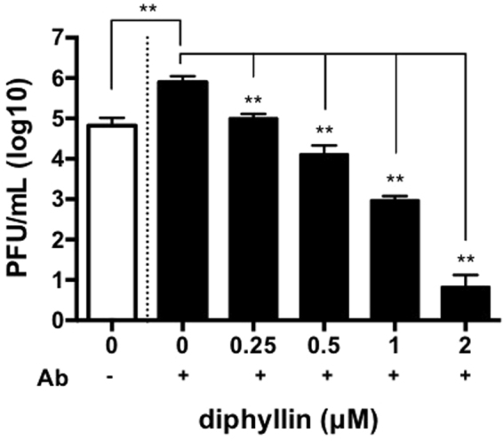
Various concentrations of diphyllin loaded PEG-PLGA nanoparticles were added to fcwf-4 cells in a direct infection (white open bar) or ADE infection (black bars) for 48 hr incubation (NTU156, MOI = 0.0035). The viral titers from the collected culture supernatants were titrated by plaques assays. Viral titers between each diphyllin-treated group and the untreated control group were compared by one-way ANOVA followed by Dunnett’s multiple comparisons test (**p < 0.01). Data in the plot present the mean ± SEM out of three replicates.