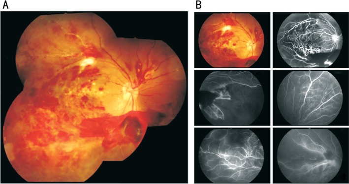
Figure 1. Fundus photograph and fluorescein angiogram of tuberculous retinal vasculitis in the right eye.
A: Retinal perivascular infiltration with branch retinal vein occlusion occurred mostly in temporal half area; B: Fluorescein angiogram demonstrated multiple large areas of capillaries dropout in the temporal side of the macula and peripheral retinal area. Early and late vascular leakage was also observed throughout the retina.

