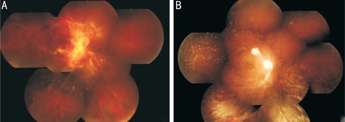
Figure 2. Fundus photographs of tuberculous retinal vasculitis in the right eye over treatment period.
A: Retinal and optic disc NV and vitreous hemorrhage occurred 2mo after treatment despite the resolution of retinal vasculitis; B: Complete regression of retinal NV after the second intravitreal bevacizumab injection was observed and there was no recurrence within a 12mo period.

