. 2017 Sep 20;18(3):359–367. doi: 10.4142/jvs.2017.18.3.359

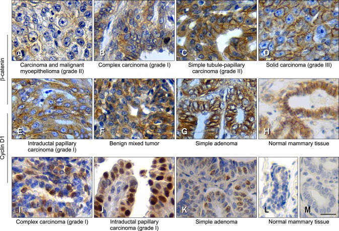
Fig. 2. The expression of β-catenin and cyclin D1 in CMTs detected via immunohistochemical staining. Tissue sections were probed with either anti-β-catenin antibodies (A–H) or anti-cyclin D1 antibodies (I–L). (A) Reduced staining of both membrane and cytoplasm; weak (D), moderate (B, E and F) and strong (C and G) staining of the cytoplasm; distinct (D), partially disappeared (C and G) or mostly disappeared (B, E and F) staining of membrane; and (H) weakly staining in cytoplasm and more than 70% of membrane staining in normal tissue section. (I–K), nuclear staining of cyclin D1. Negative control staining using PBS instead of primary antibodies was performed with normal mammary tissue (M). The red arrows indicate membrane staining of β-catenin, while the red arrowhead indicates cytoplasmic staining of β-catenin. The black arrows indicate nuclear staining of cyclin D1. The subtype and grading of the CMT samples are shown below each image. Scale bar = 20 µm.

Fig. 2