Figure-2.

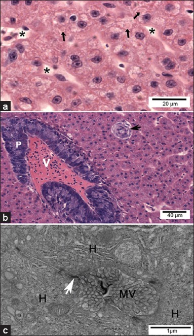
Light micrographs and transmission electron micrographs of control Pacu liver. (a) Liver parenchyma consisted mainly of hepatocytes presenting the cytoplasm voluminous with little vacuolization. Note the presence of cytoplasmic hyaline inclusions (arrows). Sinusoids are also observed (*). (b) Note the intrahepatic exocrine pancreas (P), vein (V), bile duct (arrow). (a and b) H and E. (c) Bile canaliculus formed by the plasma membranes of hepatocytes (H), where junctional complexes are noted between neighboring cells (white arrow) and the canaliculus lumen filled with hepatocyte microvillus processes).
