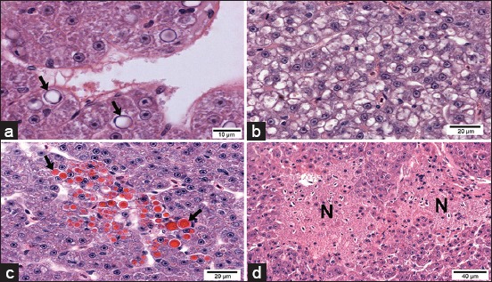
Figure-3.

Light micrographs of the liver from Pacu fingerlings exposed to 28.58 mg/L of atrazine. (a) Hepatocytes are showing nuclear vacuolization (black arrows). (b) Hepatocytes are showing cytoplasmic vacuolization. (c) The liver is showing hepatocytes with hyaline inclusions (black arrows). (d) Necrotic areas (N) (H and E).
