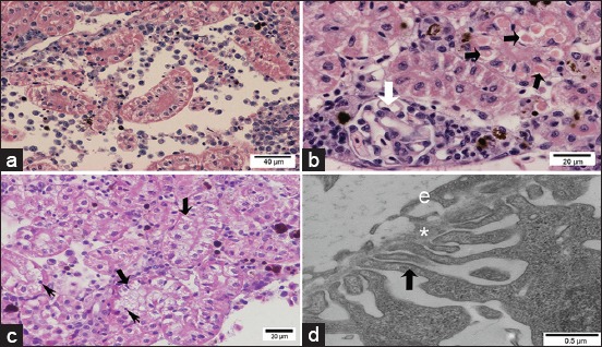
Figure-5.

Light and transmission electron micrographs of Pacu kidney from the group treated with 28.58 mg/L atrazine. (a) Area with edema and proximal tubules (PT) with vacuolated cells. (b) Proximal tubules are showing cytoplasmic hyaline inclusions (black arrows). Note the normal structure of the glomerulus (white arrow). (c) Note tubular degeneration in PT with vacuolated cells (thick arrows) and picnotic nucleus (thin arrows). (d) Glomerulus is showing podocyte pedicels (black arrow), basal lamina (*), and the endothelium of the glomerulus (e) without alterations. (a-c) H and E.
