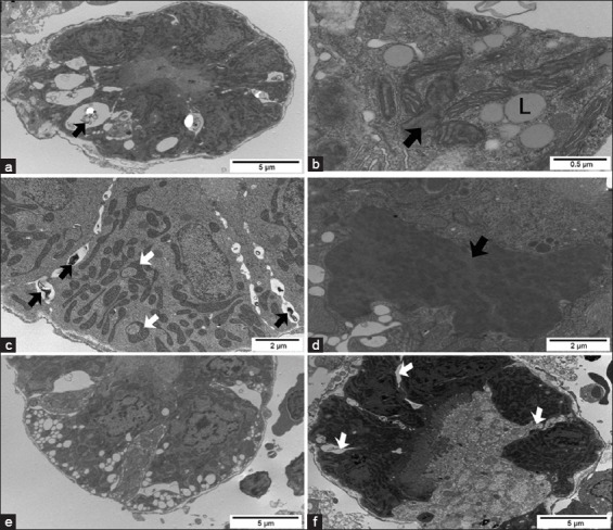
Figure-7.

Transmission electron micrographs of the kidney from Pacu treated with 28.58 mg/L of atrazine. (a) Proximal tubule (PT) cells with a large number of electron lucent vacuoles containing myelin figures (black arrow) in the basal region of the cytoplasm. (b) PT with alterations in the organization of mitochondrial cristae (black arrow) and cytoplasmic lipid inclusions (L). (c) PT with noted mitochondria with altered shape (white arrow) and enlarged intercellular space containing myelin figures (black arrow). (d) Heterogeneous inclusion (black arrow) in cells of the proximal tubule. (e) Cells of the PT showing cytoplasmic vacuolization, nuclear chromatin margination, and enlarged intercellular spaces. (f) Degenerated proximal tubule showing relatively large intercellular spaces (white arrows) in a less affected area.
