Figure-1.

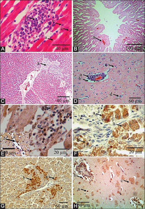
Histopathology changes and distribution of Newcastle disease virus (NDV) in internal organs of chickens from a field case in Indonesia. (A) Heart microscopic observation showed degeneration (a), mononuclear cell infiltration (b), and myocardium necrosis (c). (B) Proventriculus microscopic observation showed hyperemia on glandular epithelial cells of proventriculus glands (a). (C) Liver microscopic observation showed multifocal inflammatory cell infiltration (a). (D) Brain microscopic observation showed hyperemia with perivascular cuffing (a), edema (b), gliosis (c), and satellitosis (d). (E) Heart immunopositive reaction was detected in the cytoplasm of myocardium (a), mononuclear cell infiltration (c), and in vascular endothelial cell (insert) (b). (F) Proventriculus immunopositive reaction was distributed on glandular epithelial cells of proventriculus glands (a), and mononuclear cell infiltration (b). (G) Liver immunopositive reaction was distributed on cytoplasm of hepatocytes (a), and at the macrophage around central vein (b). (H) Brain immunopositive reaction was distributed glial cell (a), mononuclear cells of perivascular cuffing (b), and on neuron cytoplasm (c). Hematoxylin and eosin staining (A, B, C, D), immunohistochemistry staining with rabbit anti-NDV hemagglutinin-neuraminidase protein polyclonal antibody (E, F, G, H).
