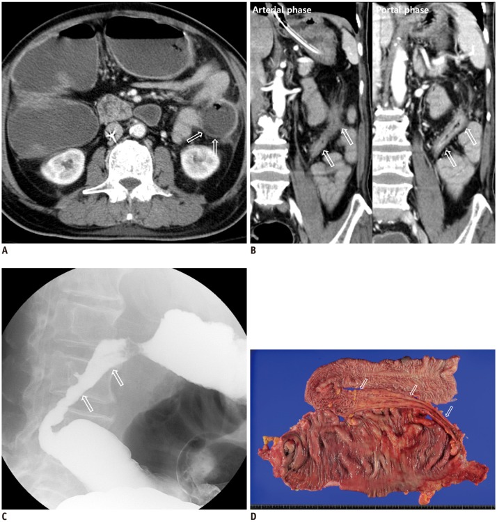
Fig. 1. 67-year-old man with multiple pre-existing and underlying pathologies (including diabetes mellitus, hypertension, chronic renal failure, and complete AV block) (patient #1) who suffered loss of consciousness during defecation.
A. CT scan performed (at time of diagnosis of ischemic bowel disease) shows some areas of decreased enhancement (arrows) in splenic flexure of colon, which is consistent with ischemic colitis. Patient had diarrhea at that time, and vital parameters were stable. Patient was managed conservatively, with antibiotics. B. Coronal arterial and portal phase images obtained 98 days after ischemic event show better bowel wall enhancement in portal phase than arterial phase (mild homogeneous enhancement in arterial phase and moderate mucosal enhancement in portal phase) (arrows) as well as vasa recta prominence around site of stricture. C. Colon study shows thickened folds (“thumb printing”), which is typical finding in cases of ischemic colitis. Patient underwent subtotal colectomy. D. Gross specimen of resected large bowel reveals approximately ten centimeters long segmental stricture (arrows) with dilatation of proximal bowel segment. This corresponds with CT image findings (arrows in B) and colon study (arrows in C). AV = atrioventricular

