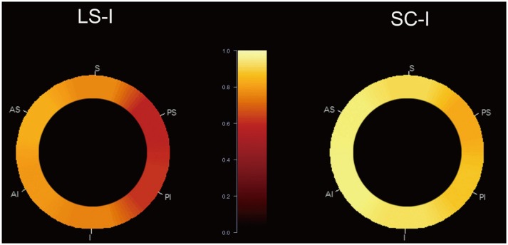
Fig. 1. Location of MCA plaque in SC-I and LS-I.
Two circular graphs show relative incidence of MCA plaque in 145 patients. MCA is segmented into 6 divisions in short-axis view. Scale 1.0 (bright yellow) refers to highest incidence of plaque (100%), and scale 0.0 (dark red) refers to lowest incidence of plaque (0%). Incidence between 0% and 100% appears in gradient color display, from maximum and minimum values. Overall incidence of MCA plaque is higher in SC-I than in LS-I, but location of plaque among 6 divisions in MCA does not differ in SC-I and LS-I. AS = antero-superior, AI = antero-inferior, I = inferior, LS-I = lenticulostriate infarction, MCA = middle cerebral artery, PI = posteroinferior, PS = postero-superior, S = superior, SC-I = striatocapsular infarction

