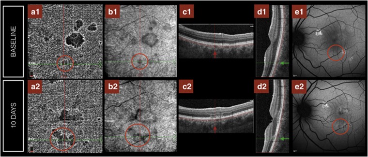
Figure 3.
Evolution of Phase 1 lesion to a Phase 2 lesion and consecutive stages (left eye of Patient 3). The lesion is identified with a red circle. OCT-A (a1) discloses a small area of hypoperfusion, equally en face-OCT shows mild hyporeflectivity at the level of the choriocapillaris (b1), but FAF and SD-OCT are essentially normal in the early stage (c1–e1). This choriocapillaris hypoperfusion identified on OCT-A had increased in size 10 days later (a2) also visible in the en face-OCT (b2) with the lesion now identifiable on the SD-OCT in the horizontal (c2, red line) and vertical B-scans (d2, green line), as well as with FAF (e2). A full color version of this figure is available at the Eye journal online.

