Figure 2.

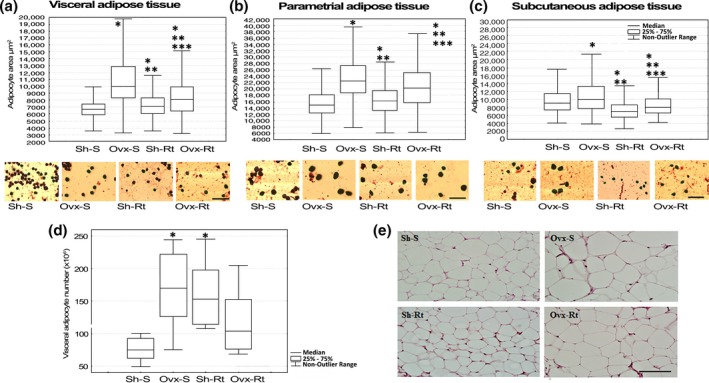
Adipocyte area and adipocyte number. Adipocyte area measured by osmium tetroxide fixation and photomicrographs of corresponding group. (a) Visceral adipose tissue; (b) parametrial adipose tissue; (c) subcutaneous adipose tissue. Representative adipocytes photomicrographs (10 μm – original magnification ×400). Results are expressed as means and standard deviation of the mean (n = 8 per group). At least 400 adipocytes of each depot fat per animal had their mean diameter measured. (d) Adipocyte number measured by haematoxylin–eosin (HE). (e) Visceral adipose tissue histology. Slides were randomly digitized under a light microscope for HE (10 fields per animal – 20 μm, original magnification ×400). Different superscripts: * denotes significantly different from sham sedentary; ** denotes significantly different from ovariectomy; *** denotes significantly different from resistance training. Groups are significantly different from each other at P < 0.05. Abbreviations: Sh‐S, sham sedentary; Ovx‐S, ovariectomized sedentary; Sh‐Rt, sham resistance training; Ovx‐Rt, ovariectomized resistance training. [Colour figure can be viewed at wileyonlinelibrary.com].
