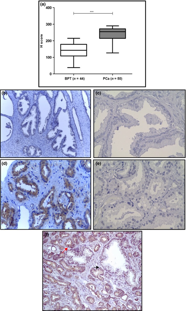
Figure 2.

CXXC5 protein expression in benign prostate tissue and prostate cancer. (a) CXXC5 protein expression was evaluated by IHC and compared in prostate tissue with benign glands (n = 44), and prostate tissue with tumour glands (n = 50). Immunohistochemistry staining of CXXC5 in benign prostate tissue with absent to mild staining (b) and prostate cancer tissue with strong staining (d). Negative control (no primary antibody) for benign prostate tissue (c) and malignant prostate tissue (e). CXXC5 staining in malignant acini (red arrow) compared to benign acini (black arrow) from the same patient (f). Magnification: 100×. Brown colour: DAB. Blue colour: Haematoxylin counterstain. ***P < 0.0001. [Colour figure can be viewed at wileyonlinelibrary.com].
