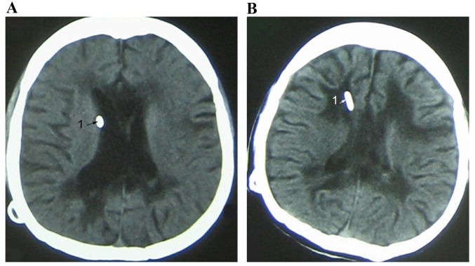
Figure 4.
Ventricle images of a female patient with SVS (age 22). (A) Imaging indicated that the ventricle end of the shunt was in close contact with the ventricle wall (arrow). (B) Paraventricular edema was caused by chronic ventricular infection after VP shunting and the ventricle was in slit state (arrow). SVS, slit ventricle syndrome; VP, ventriculoperitoneal.

