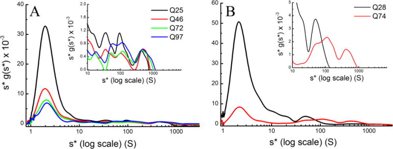
Figure 3.

MSM–WDA analysis of (A) HttQ25-eGFP, HttQ46-eGFP, HttQ72-eGFP, and HttQ97-eGFP in D. melanogaster and (B) GFP-HttQ28 and GFP-HttQ74 in C. elegans. All samples prepared in lysis buffer were centrifuged at 3000, 6000, 10000, 20000, 30000, and 50000 rpm until the meniscus was cleared. The log plot of s*g(s*) vs s* shows the complete distribution of s values ranging from 0.8 to 3000 S, and the inset focuses on the 20–3000 S region to better highlight the distribution of the intermediate aggregates.
