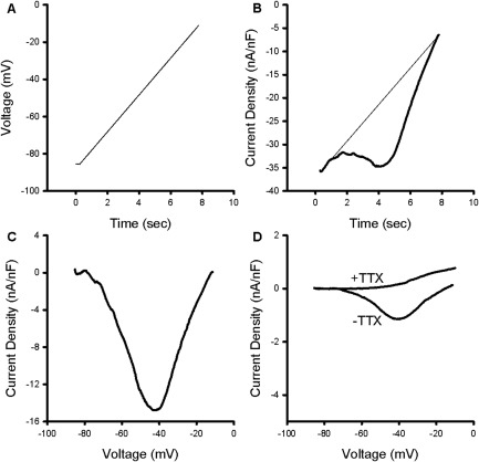
Figure 2.

Characterization of a tetrodotoxin (TTX)‐sensitive persistent inward current in ClC muscle. (A) The voltage protocol used to identify persistent inward currents (PICs). From a holding potential of −85mV, fibers were depolarized to −10mV at a rate of 10mV/s. (B) The current trace generated by the ramp depolarization in normal K+ solution with 20μM nifedipine. A fit line (thin) is drawn using the first 0.5 seconds of the raw trace, representing the leak current. Deviations from the leak current/fit line in the negative direction are consistent with activation of a PIC. (C) Shown is a trace generated by subtracting the leak trace shown in B and plotting against voltage. (D) Plot of leak‐subtracted PIC in K+‐free solutions (−TTX). The PIC was blocked by the addition of 1μM TTX to the external solution (+TTX).
