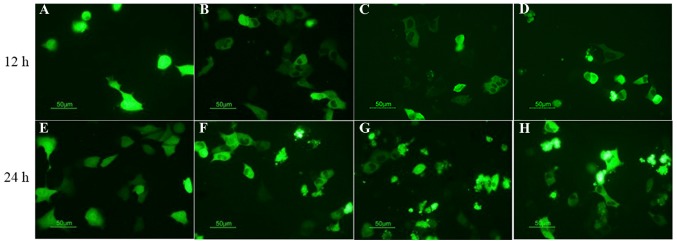
Figure 3.
Fluorescence microscopy analysis of the apoptotic behavior of CRT-MUC1-coated 4T1 cells. pEGFP-MUC1-CRT was transiently transfected into 4T1 cells and treated with (A) 0 µg/ml, (B) 2 µg/ml, (C) 4 µg/ml and (D) 8 µg/ml mitoxantrone for 12 h, or with (E) 0 µg/ml, (F) 2 µg/ml, (G) 4 µg/ml and (H) 8 µg/ml mitoxantrone for 24 h and the results were observed by fluorescence microscopy. Fluorescence microscopy was used to detect that the apoptotic behavior of CRT-MUC1-coated 4T1 cells treated with mitoxantrone was dose- and time-dependent. Magnification, ×400. MUC1, mucin 1; CRT, calreticulin.

