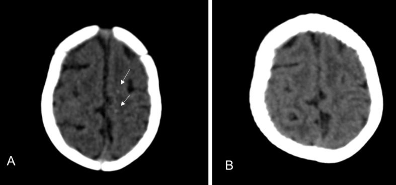
Fig 3 CT scans of only child with congenital Zika syndrome whose cerebral calcifications were no longer visible. Initial scan (A) shows tenuous punctate calcifications at the cortical-white matter junction in frontal lobes (arrows). (B) Calcifications are no longer visible at one year follow-up
