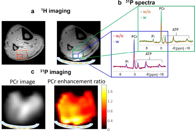
Figure 5.
In vivo results. (a) 1H images with and without the metamaterial. (b) 31P spectra of two size voxels (single voxel with maximal enhancement and four-combined voxels) with and without the metamaterial. (c) Phosphocreatine (PCr) image with the metamaterial in place and a map of the enhancement ratio with the metamaterial compared to without the metamaterial. The maximum enhancements (for the same excitation tip angle) were 1.8 and 2.1 for 31P and 1H, respectively. A schematic cross-sectional overlay of the metamaterial (white) is shown in the images.

