Fig. 1.

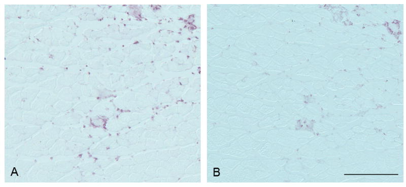
Anti-7/4 immunostaining (A) showed medium intensity and was observed in connective tissue and myofibers. Anti-Ly6C/G immunostaining (B) was less intense and was located mainly within myofibers. Bar = 200 μm.

Anti-7/4 immunostaining (A) showed medium intensity and was observed in connective tissue and myofibers. Anti-Ly6C/G immunostaining (B) was less intense and was located mainly within myofibers. Bar = 200 μm.