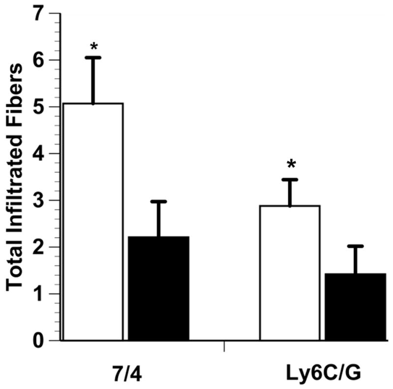
Fig. 2.

7/4-Positive and Ly6C/G-positive myofibers in injured lateral gastrocnemius muscle. White bars, E+ mice; black bars, E-mice. E+ mice had a significantly larger number of 7/4-positive and Ly6G-positive myofibers than E- mice (*p < 0.05). Total infiltrated fiber is the absolute number of stained fibers in the analyzed area of interest and data are means ± SE.
