
Figure 4. NBS1S432 phosphorylation is cell cycle regulated and required to protect newly replicated telomeres.
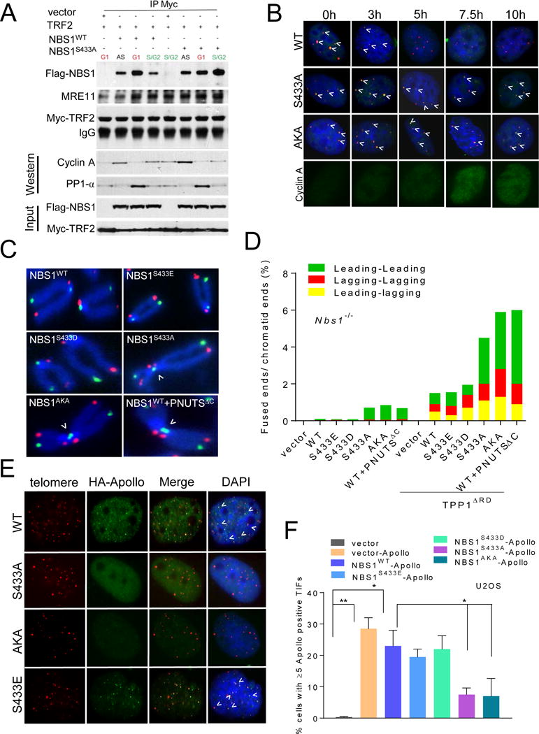
A. Fucci U2OS cells transfected with indicated DNA constructs were FACS sorted to purify G1 and S/G2 cells. Sorted cells were immunoprecipated with anti-Myc antibody and then immunoblotted with anti-Myc and anti-Flag antibodies. Inputs represent 5% of the total cell lysate used for the IP. Cyclin A was used to mark S/G2 phase of the cell cycle. B. U2OS cells synchronized with 2mM thymidine and 1.0 μg/ml aphidicolin were fixed, telomeres visualized by PNA-FISH (red), NBS1 visualized by anti-Flag antibody (green), and DAPI (blue). Anti-cyclin A antibody was used to mark S/G2. C. Metaphases prepared from Nbs1−/− MEFs reconstituted with WT mNBS1, mNBS1S433 mutants or PNUTSΔC were analyzed by CO-FISH. FITC-OO-(TTAGGG)4 (green, leading strand), Tam-OO-(CCCTAA)4 (red, lagging strand) and DAPI for chromosomes (blue). Arrowheads indicate leading-leading chromatid fusions. D. Quantification of chromatid fusions observed in (C). E. Localization of Apollo/SNM1B in U2OS cells expressing WT Flag-mNBS1 or Flag-mNBS1S433 mutants. Telomeres were visualized with PNA-FISH (red), anti-HA antibody to visualize Apollo/SNM1B (green), and DAPI (blue). F. Quantification of Apollo/SNM1B foci in (E). Data represents the mean of three independent experiments ± SEM; n>200 nuclei scored per experiment. *: p<0.01; one-way Anova. See also Figures S3 and S4.