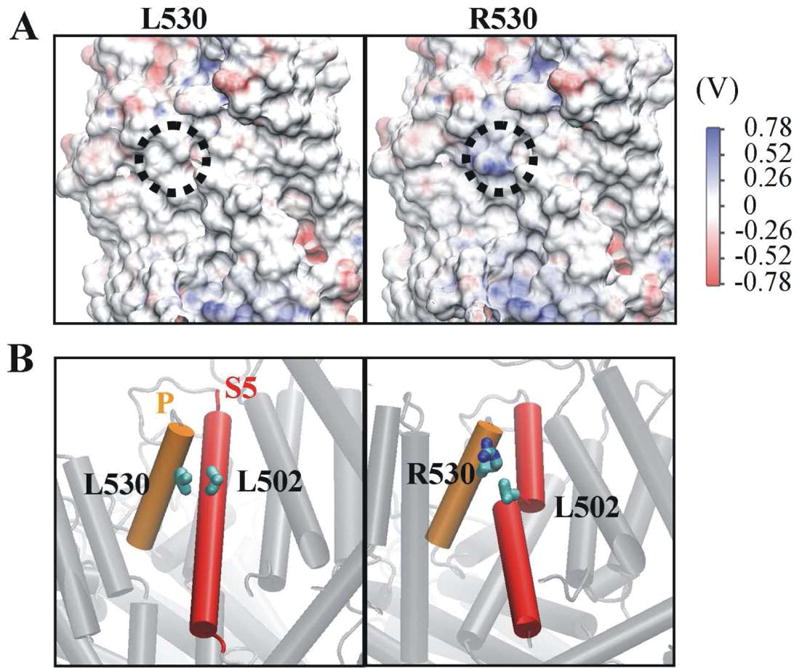
Fig. 2. The L530R variation changes electrostatic potential at position 530 and damages the secondary structure of transmembrane helix 5.

(A) The electrostatic potential of residue 530 (dotted circle) shifted positively when L530 was replaced by R530. (B) Transmembrane helix 5 (S5) broke into two helices in the R530 variant as a result of the disruption of the interaction between R530 and L502. The pore helix (P) and S5 are shown in orange and red, respectively. The carbon and nitrogen atoms of residue 530 are labeled in cyan and blue, respectively. For clarity, the hydrogen atoms of residue 530 are not shown.
