
Figure 2.
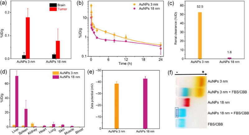
(a) Distribution of AuNPs with different sizes in gliomas and normal brain tissues at 24 h p.i. (n = 3). Before collecting the organs, AuNPs in the bloodstream were removed by perfusion. (b) Pharmacokinetics, (c) renal clearance efficiency, and (d) biodistribution of AuNPs with different sizes at 24 h p.i. (n = 3). (e) Zeta potential values of 3-nm and 18-nm AuNPs in PBS at pH 7.4. (f) Serum binding test of 3-nm AuNPs and 18-nm AuNPs by gel electrophoresis. The dark blue color in the rectangle was derived from serum binding and aggregation of 18-nm AuNPs. CBB, Coomassie brilliant blue dye was added to stain FBS. AuNPs were incubated with 10% (v/v) FBS in PBS at 37 °C for 1 h.