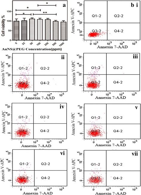
Fig. 3.

Cell viability of neuroglia incubated with different concentrations of AuNS@PEG nanoparticles for 24 h (a); the apoptosis of cells induced by AuNS@PEG nanoparticles is shown by flow cytometry: control (bi), 25 ppm (ii), 50 ppm (iii), 100 ppm (iv), 200 ppm (v), 500 ppm (vi), and 1000 ppm (vii)
