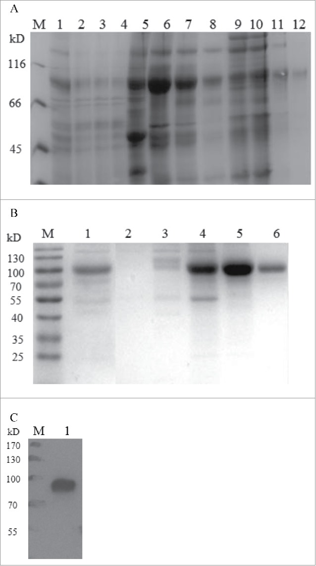
Figure 4.

Purification of recombinant human HSA-eTGFBR2. (A) SDS-PAGE analysis of the fractions separated by Blue Sepharose column. M: marker; lane 1: the supernatants of fed-batch culture; lane 2–4: flow through; lane 5: fractions eluted by 30% buffer B; lane 6–8: fractions eluted by 100% buffer B; lane 9–12: fractions eluted by 1M Arg. (B) SDS-PAGE analysis of the fractions separated by Q Sepharose column. M: marker; lane 1: loading; lane 2: flow through; lane 3: washing fractions; lane 4–6: eluting fractions. (C) Western blot analysis of purified HSA-eTGFBR2 fusion protein.
