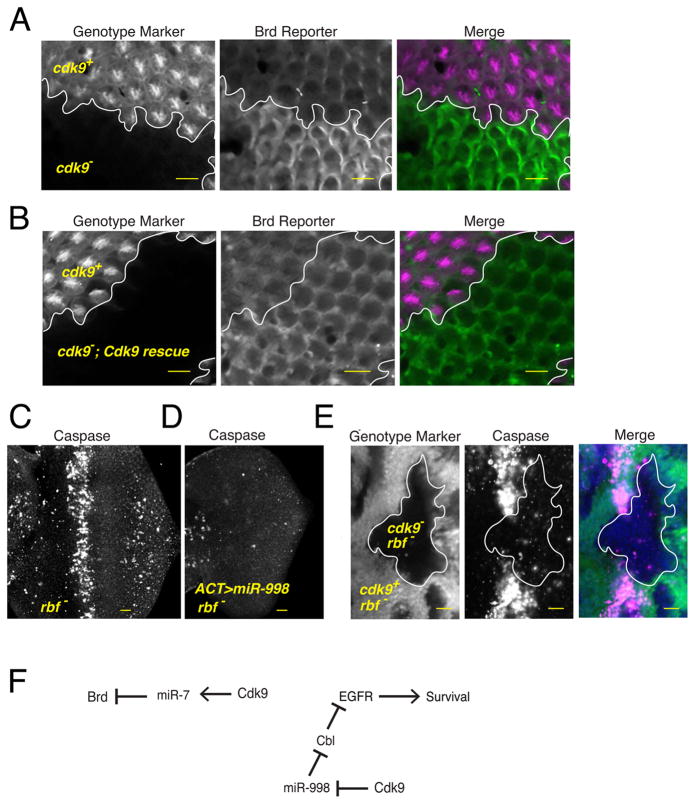
Figure 6. Differential processing has a significant effect on gene silencing in vivo.
See also Figure S5. (A,B) Expression of the Brd reporter gene in the developing eye. Each image contains approximately 800 cells, some of which are genetically wildtype and some of which are mutant for the cdk9 gene. Cell genotypes have been marked by the presence or absence of an RFP marker, as indicated. RFP and GFP channels are shown separately along with a merged image. Since cdk9 mutant cells in (B) also expressed the Cdk9 rescue transgene, silencing of the Brd reporter is restored within these cells. (C) When rbf mutant eye discs are stained for activated caspase protein, they show a zone of prevalent cell apoptosis within the morphogenetic furrow. (D) This zone is absent if eye cells overexpress miR-998 via the GAL4/UAS system. (E) When rbf mutant cells are also mutant for cdk9, then there is greatly reduced apoptosis. (F) A summary of the genetic experiments showing the pathways of gene regulation downstream of Cdk9. Scale bars for A–E, 10 μm.

