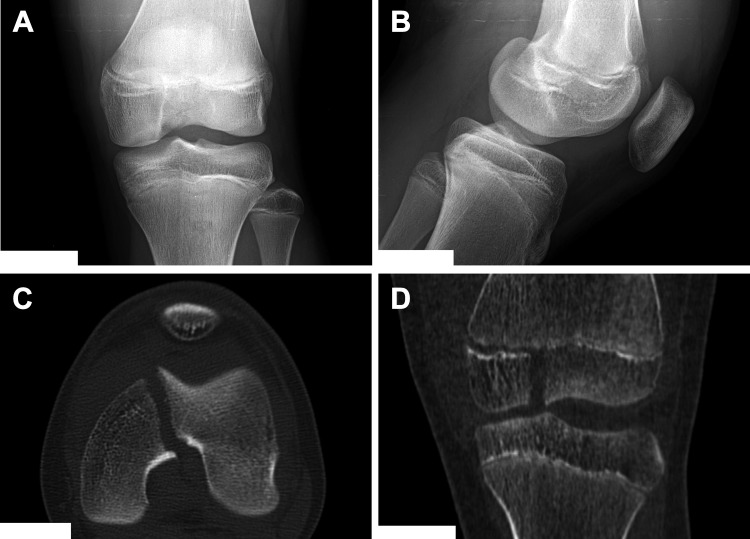
Figure 2.
(A, B) Anteroposterior and lateral radiographs of an adolescent male football player with no obvious fracture visualized on plain radiographs but large effusion best seen in the suprapatellar pouch on the lateral view. (C, D) Coronal and axial computed tomography images confirming the intra-articular fracture and demonstrating how marked displacement can be underrecognized on plain films. Image courtesy of SD PedsOrtho.

