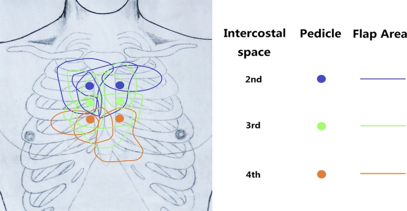
FIGURE 2.

After keloid resection, the recipient vessels were explored and identified in the second, third and fourth intercostal spaces. The SCIP flaps were transplanted and anastomized with the appropriate vessels regarding the inset position and vessel caliber. The sketch shows the dimensions of the SCIP flaps and their intercostal perforator pedicles.
