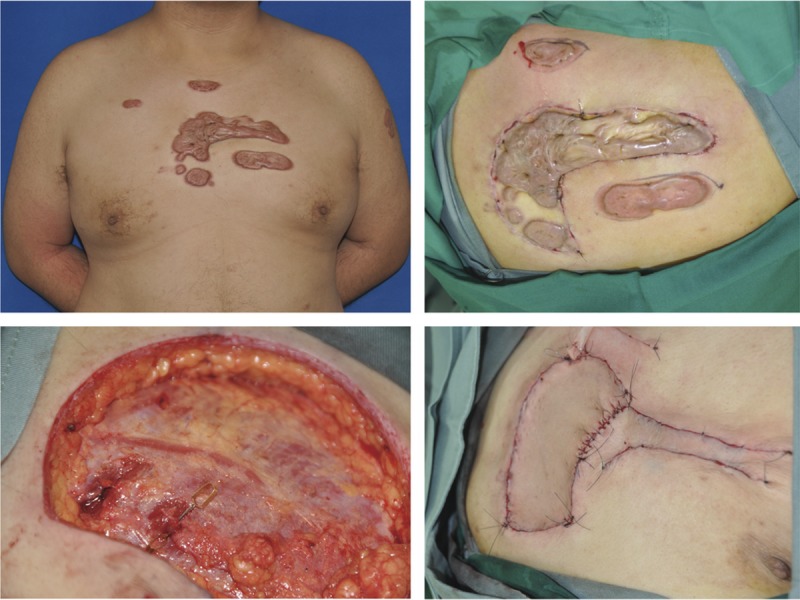
FIGURE 3.

This patient had a tendency to develop numerous keloids in the presternal area and upper arm (Left above). He received the precut procedure on the first day, in which the biggest presternal keloid was incised peripherally, and intra-dermal sutures were used to close the wound (right above). On the third day, the keloid was removed surgically, and the second intercostal perforator was identified above the deep fascia (left below). A SCIP flap was transplanted for the coverage of the major per-sternal defect, while the minor defects were closed using advancement local flaps (right below).
