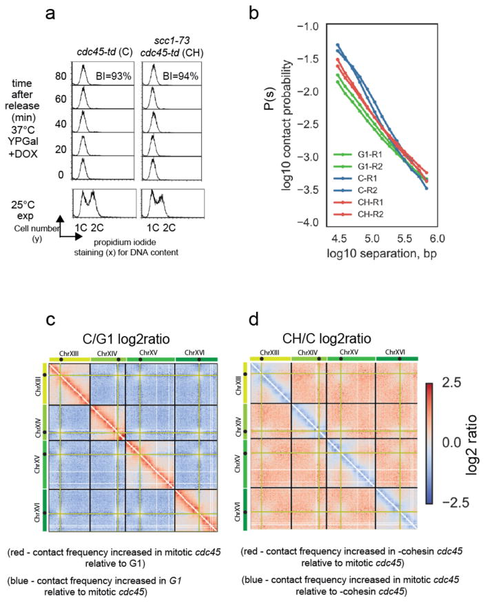
Figure 4. Mitotic cohesin-dependent conformational changes are independent of sister chromatid cohesion.
a) FACS analysis of DNA content and budding analysis of cdc45-td (C) and cdc45-td scc1-73 (CH) cells following release from G1 arrest into a nocodazole enforced mitotic block. Budding index (BI) confirmed that mitotic cells had activated CDK while FACS of DNA stained cells confirmed no DNA replication has taken place. Representative images shown from one of two independent experiments comparing C to CH.
b) Contact probability, P(s) versus genomic separation, s, for Hi-C of mitotic cdc45-td (C) mitotic cdc45-td scc1-73 (CH), and wt G1 cells (G1). The P(s) from each of the two independent experiments for each condition are shown.
c) Log2 ratio of C (cdc45 depleted cells arrested in mitosis with nocodazole) contact dataset over G1 dataset (C/G1). Contact maps for ratio plot were assembled from two independent experiments for each condition.
d) Log2 ratio of –cohesin C dataset over C dataset (CH/C).

