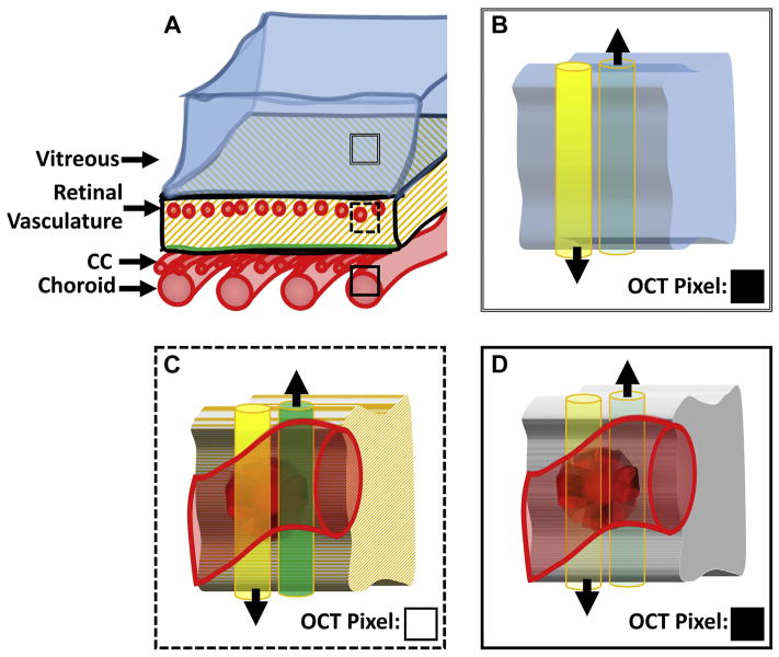
Figure 3.
Illustration of the different causes of low OCT signal. A, Sketch of the posterior eye, from the vitreous to the choroid. The 3 different boxes— double-lined, dashed, and solid—correspond to panels B, C, and D, respectively. For each of B, C, and D, the yellow (left) cylinder corresponds to the incident OCT beam and the green (right) cylinder corresponds to the backscattered light. These cylinders are shown side-by-side for visual clarity; however, in reality they are superimposed. The black arrows point in the direction of energy (photon) propagation. More opaque yellow coloring (B and C) corresponds to a stronger (i.e., less attenuated) incident OCT beam; more transparent yellow coloring (D) corresponds to a weaker (i.e., more attenuated) incident beam. Similarly, more opaque green coloring, C, corresponds to stronger backscattered light (i.e., more backscattered photons); more transparent green coloring, B and D, corresponds to weaker backscattered light (i.e., less backscattered photons). The squares in the bottom right corner of B–D, labeled “OCT Pixel,” indicate the hypothetical value of the OCT pixel corresponding to that tissue location, with black being low and white being high. CC = choriocapillaris. B, Tissue cube from the vitreous humor. The incident beam is strong, having been minimally attenuated. However, because the vitreous humor does not contain backscatterers, the backscattered light is small. Thus, the corresponding OCT pixel is black. C, Tissue cube from a layer intersecting the retinal vasculature. The incident beam is still strong, and due to the presence of backscattering tissue (in this case an erythrocyte), there is significant backscattered light. Thus, the corresponding OCT pixel is white. D, Tissue cube from the choroid/choriocapillaris. The incident beam is strongly attenuated, having passed through the retinal pigment epithelium and potentially some choroidal vasculature. Thus, even though the tissue at this location contains back-scatterers, the backscattered light is minimal. Thus the corresponding OCT pixel is black.

