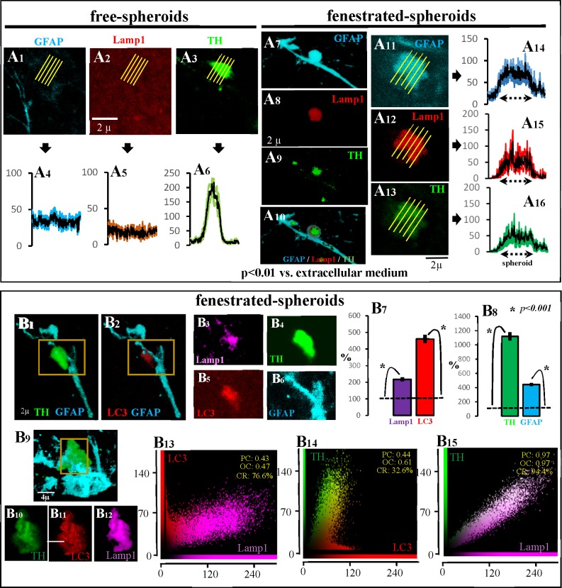
Fig 4. Main characteristics of fenestrated-spheroids.
A1-A6 show an example of the absence of Lamp1 (A2) in free-spheroids which present TH (A3 and A6) but not GFAP (A1 and A4) immunoreactivity. Figs A7-A10 show an example of a fenestrated-spheroid (A10) showing TH (A9), GFAP (A7) and Lamp1 (A8) immunoreactivity (this example is also shown in S6–S9 Animations). Figs A11-A16 show another example of a fenestrated-spheroid showing TH (A13), GFAP (A11) and Lamp1 (A12) immunoreactivity (the immunoreactivity distribution within spheroids is shown in A16, A14, and A15, respectively). The fenestrated-spheroids (B4/B6 present an amplification of the yellow square of B1) which showed high LC3 immunoreactivity (B2/B5) also showed the lysosomal marker Lamp1 (B3). Thus, LC3 and Lamp1 immunoreactivity (B7) was higher in fenestrated spheroids (B8) than in the surrounding extracellular space (B7 and B8 show the mean ± standard error of values normalized according to the extracellular immunoreactivity). B10-B12 show an amplification of the yellow square shown in B9. The LC3 (B11) and Lamp1 (B12) observed in fenestrated-spheroids (B9/B10) showed co-localization inside the spheroid (B13; PC: Pearson’s correlation; OC: overlap coefficient; CR: co-localization rate), suggesting the production of autophagolysosomes in the fenestrated-spheroids. The findings of TH-LC3 (B14) and of TH-Lamp1 (B15) co-localization suggest that DAergic debris is being stored in autophagolysosomes.

