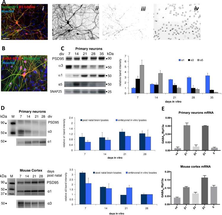
Fig 1. Biochemical characterization of cortical network characteristics and GABAAR subunit expression in primary cortical neurons and in mouse frontal cortex tissue.
A. Immunocytochemistry characterization of the primary cortical neurons (28 DIV). i. Staining with antibodies for synapsin-1 (green), β3-tubulin (red) and Hoechst-Bisbenzimid (blue). ii. Neurons. iii. Isolated synaptic punctae. iv. Nuclei. B. Immunocytochemistry characterization of α1 GABAAR protein expression in the primary cortical neurons (28 DIV). Staining for α1 (red), β3-tubulin (green) and Hoechst-Bisbenzimid (blue). C. Western blot analysis of α1, α3 and α5 GABAAR protein expression levels in the primary neurons (7, 14, 21, 28 and 35 DIV). D. Western blot analysis of α1 and α3 protein expression levels in the primary neurons (7, 14, 21 and 28 DIV) and in postnatal mouse frontal cortex (7, 14, 21 and 28 days postnatally). E. RT-qPCR analysis of α4, β1, β2, β3 and δ mRNA expression levels (± S.D.) in the primary neurons and in postnatal mouse cortex tissue (relative to the expression of the reference gene RPL13a).

