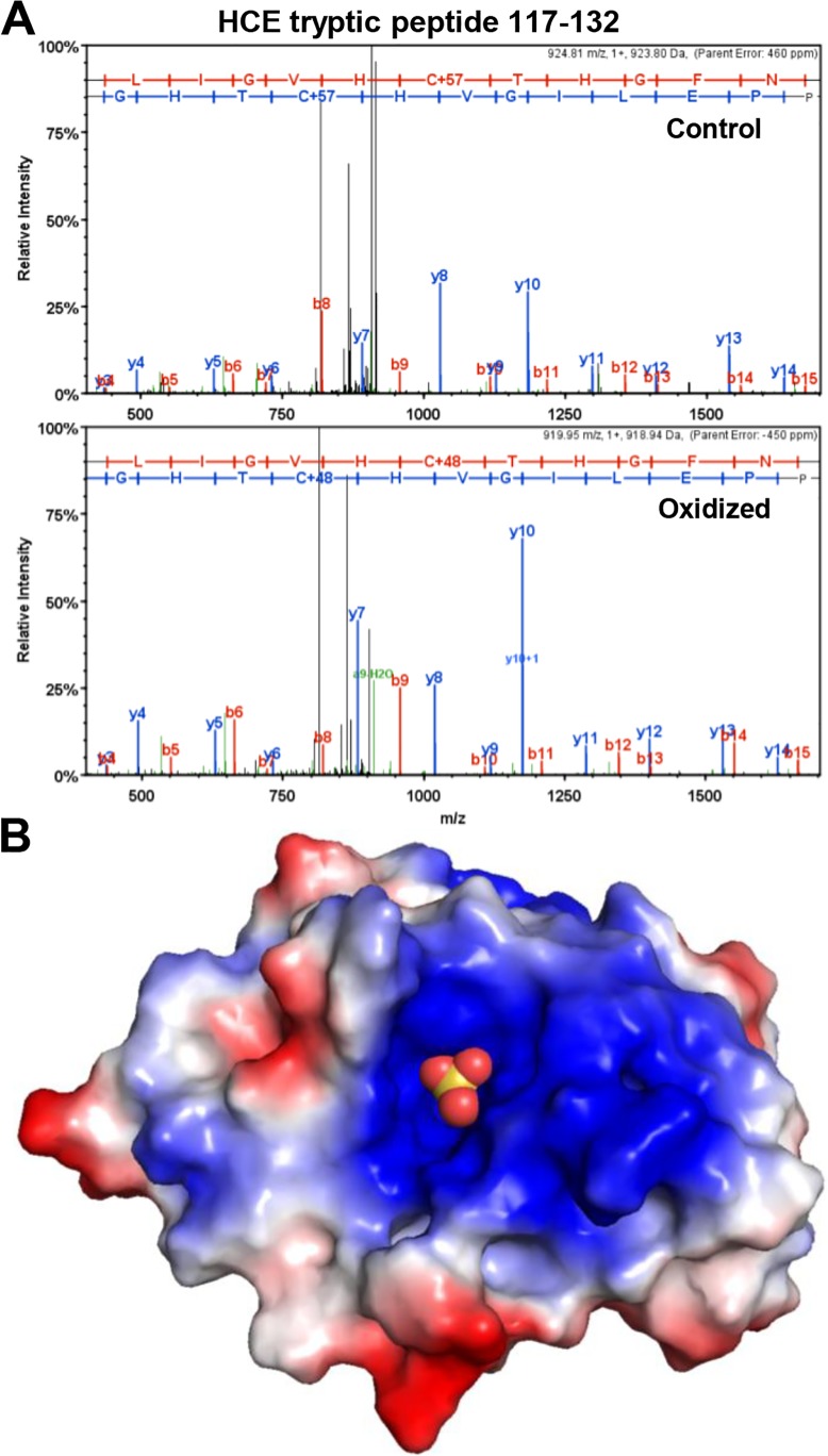
Fig 3. Cys126 is the site of oxidation in HCE.
A, Spectra from MS/MS fragmentation analysis of tryptic peptide (aa 117–132) from HCE treated with DTT (Control) or 0.6 mM H2O2 and then quenched with 10-fold molar excess DTT (Oxidized). B, Model of HCE triphosphatase domain with active site Cys126 oxidized to sulfonic acid (balls). Model was built in PyMol using mouse triphosphatase domain oxidized with tungstate treatment (sulfonic acid group added) PDB 1I9T [20]. Positive (blue) and negative (red) charged regions are indicated.

