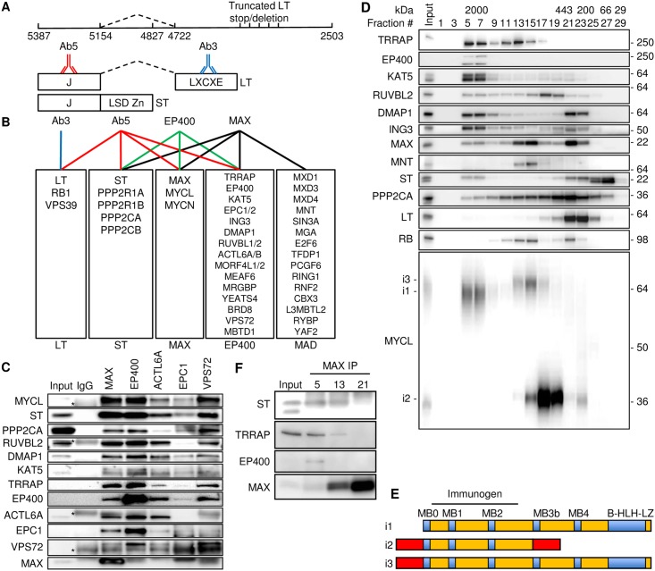
Fig 1. MCPyV ST binds MYCL and EP400 complex.
(A) MCPyV early region showing nucleotide positions for LT start (5387), ST stop (4827), LT stop (2503), and LT splice donor (5154) and acceptor (4722) and approximate positions of mutations that result in truncated LT found in MCC. LT and ST share an N-terminal J domain. The ST unique domain contains the LSD and Zn fingers. LT splices from J domain to a second exon containing the LXCXE or RB1 binding motif. Antibody Ab3 binds LT only and Ab5 binds both LT and ST. (B) Identification of co-precipitating proteins by MudPIT with antibodies Ab3 (LT, blue), Ab5 (LT/ST, red), EP400 (green) and MAX (black). See S1 Table for details. (C) MKL-1 lysates were immunoprecipitated (IP) with indicated antibodies (top) followed by immunoblotting with indicated antibodies (left). Asterisks indicate non-specific bands in IgG control immunoprecipitation lane. (D) MKL-1 lysates (Input) were separated in a Superose 6 column and fractions (#) were blotted with antibodies indicated on left. Protein size markers in kDa indicated at top and right. (E) Three MYCL isoforms (i1, i2, i3) are indicated (see also S1 Fig). Immunogen of MYCL antibody contained MYCL-i1 residues 16–139. (F) Fractions #5, 13 and 21 from Fig 1D were immunoprecipitated with MAX antibody and blotted.

