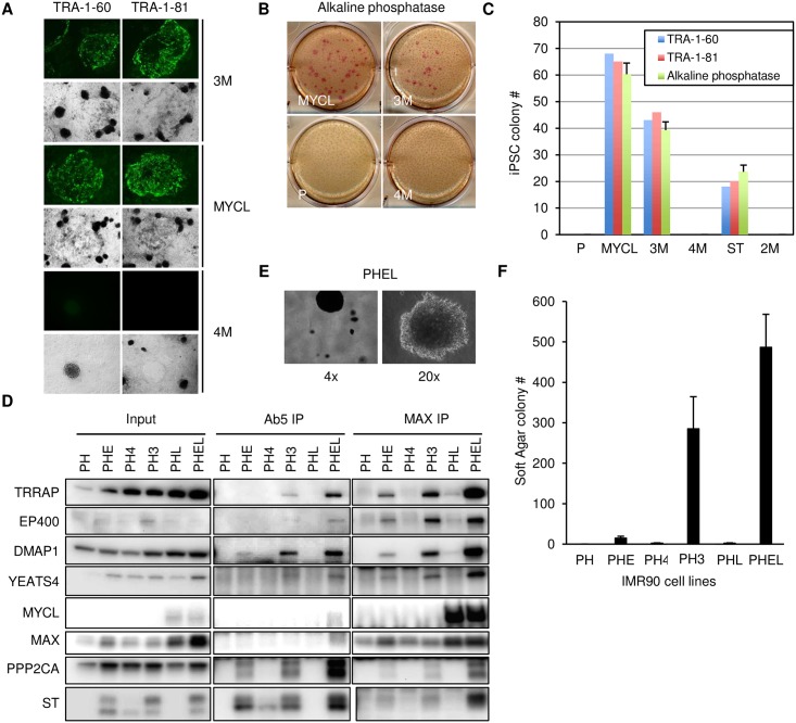
Fig 5. MCPyV ST, MYCL and EP400 complex cooperate to reprogram and transform cells.
(A) HFK-hTERT cells were transduced with Dox-inducible OCT4, SOX2 and KLF4 (P) and stably expressed MYCL, 3M or 4M MCPyV ST. Cells were treated with Dox for 31 days and then were immunostained with fluorescent antibodies to TRA-1-60 or TRA-1-81. Light field images demonstrate flat iPSC colonies formed with 3M and MYCL but not from 4M. (B) Cells were stained with alkaline phosphatase one day after immunostaining (Fig 5A). (C) Number of iPSC colonies detected after 31 days. Three biological replicas were performed. Data are presented as mean (SD). (D) IMR90 cells stably expressing dominant negative p53 and hTERT (PH) were transduced with MYCL (PHL) or tumor derived MCPyV ER region containing truncated LT and wild type ST (PHE) and MYCL (PHEL) or 3M mutant ST (PH3) and 4M mutant ST (PH4). Lysates (Input) were prepared from indicated cells, immunoprecipitated with Ab5 or MAX antibodies followed by immunoblotting with the indicated antibodies. (E) Images of soft agar colonies from PHEL cells (4X or 20X magnification). (F) Anchorage independent growth of IMR90 cells indicated in D (105 cells) plated in soft agar and cultured for 4 weeks. Three biological replicas were performed. Data are presented as mean (SD).

