
Fig. 3.
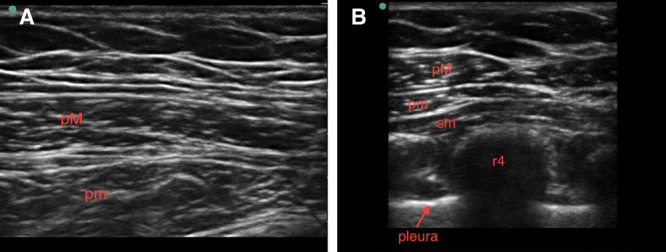
Ultrasound images of Pecs I (A) and Pecs II (B). Pecs I and II, pectoral nerve block types I and II; pM, pec major; pm, pec minor; r4, rib 4; sm, serratus muscle. Images courtesy of Jacob Hutchins, MD.
Official websites use .gov
A
.gov website belongs to an official
government organization in the United States.
Secure .gov websites use HTTPS
A lock (
) or https:// means you've safely
connected to the .gov website. Share sensitive
information only on official, secure websites.
Ultrasound images of Pecs I (A) and Pecs II (B). Pecs I and II, pectoral nerve block types I and II; pM, pec major; pm, pec minor; r4, rib 4; sm, serratus muscle. Images courtesy of Jacob Hutchins, MD.