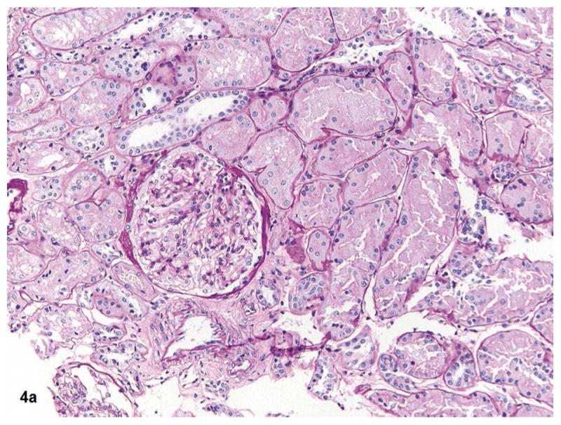
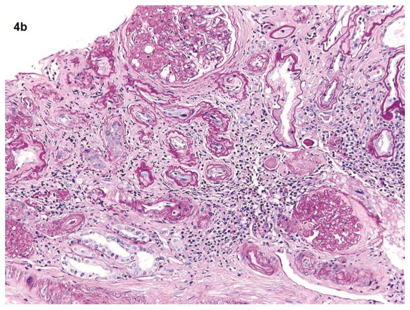
Fig. 4a–b.


Fig 4a shows the kidney biopsy pathology from a 25 years old female with proteinuria for 3 months and normal kidney function (eGFR >60). The renal cortex shows glomerulus, tubules, and vessels within no significant global glomerulosclerosis, interstitial fibrosis, or vascular sclerosis (PAS 20x). Total kidney pathology score for this subject is 0. Fig 4b shows the kidney biopsy pathology from a 62-year-old male with long term type 2 diabetes mellitus and hypertension. His eGFR is 34. Renal cortex with severe (grade 3) interstitial fibrosis, tubular atrophy, chronic interstitial inflammation, global glomerulosclerosis and vascular sclerosis (PAS 20x). His kidney biopsy pathology result indicates a pathologic severe CKD.
