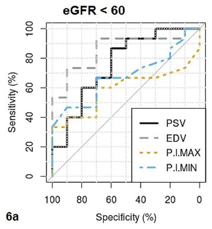
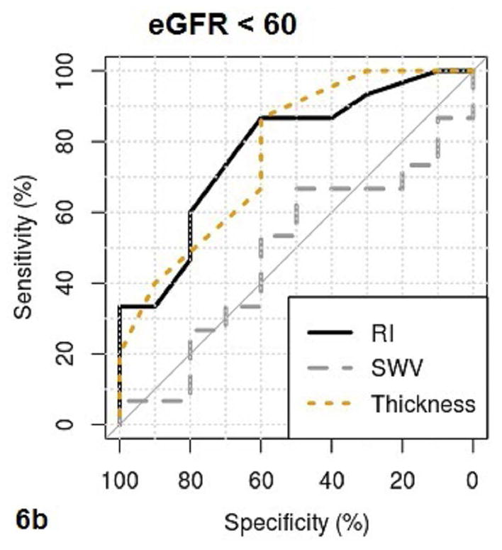
Fig. 6a–b.


These figures show the area under the receiver operating characteristics (AUROC) of QUI parameters of PSV, EDV, maximal pixel-intensity (P.I.Max), minimal pixel-intensity (P.I.Min) (6a), and RI, SWV, cortical thickness (Thickness) (6b) for determining CKD with eGFR <60. The cutoff value, sensitivity and specificity of each QUI parameter in determining eGFR <60 are listed in Table 5.
