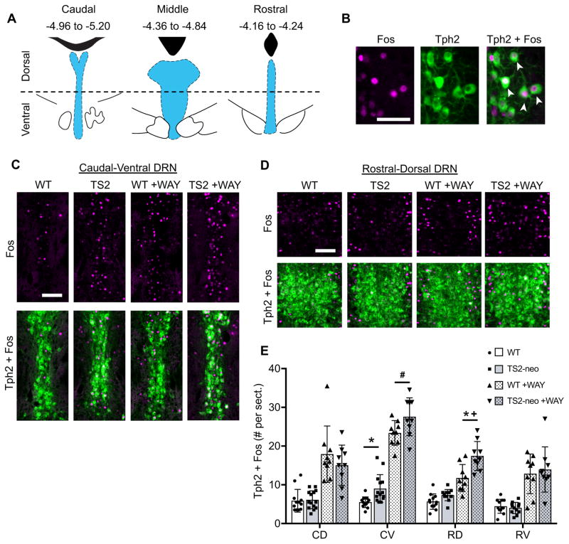
Figure 4.
Dorsal raphe nuclei (DRN) 5-HT neuron activity and 5-HT1A dependent feedback inhibition following forced swim in WT and TS2-neo mice. A, Location of the DRN (blue) relative to the 4th ventricle or cerebral aqueduct (black), and bregma coordinates of DRN subregions delineated across the rostral-caudal and dorsal-ventral axes. B, Representative DRN neurons immunofluorescently labeled for Fos (magenta, left) and Tph2 (green, middle). White arrows in the merged image (right) indicate double-labeled cells. Scale bar, 50μm. C–D, Immunofluorescent labeling of Fos (top) and merged images of Fos + Tph2 (bottom), with and without administration of 5-HT1A antagonist WAY-100635 (WAY), Scale bars, 100μm. E, Average # of Fos+Tph2 cells counted per tissue section from DRN subregions, ±SD. CD = caudal-dorsal, CV = caudal-ventral, RD = rostral-dorsal, RV = rostral-ventral. *, FDR-adjusted q < .05 between TS2-neo and corresponding WT control group. #, p < .05 and q > .05 between TS2-neo and corresponding WT control. +, genotype by drug interaction p < .05.

