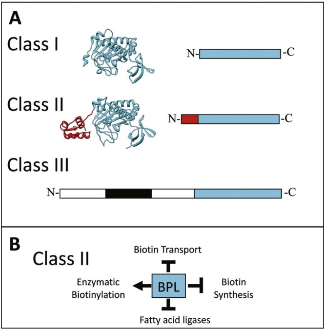
Fig. 2.

Biotin Protein Ligase. (A) The relative sizes of the three structural classes of BPLs are shown. The conserved catalytic region is depicted in blue, the DNA binding domain of Class II enzymes in red and the proof reading domain in human BPL is boxed black.17, 18 The structures of BPLs from M. tuberculosis [PDB 3RUX19] and E. coli [PDB 2EWN20] are highlighted. (B) Schematic overview showing the single protein model of protein biotinylation and transcriptional regulation in Class II BPLs.
