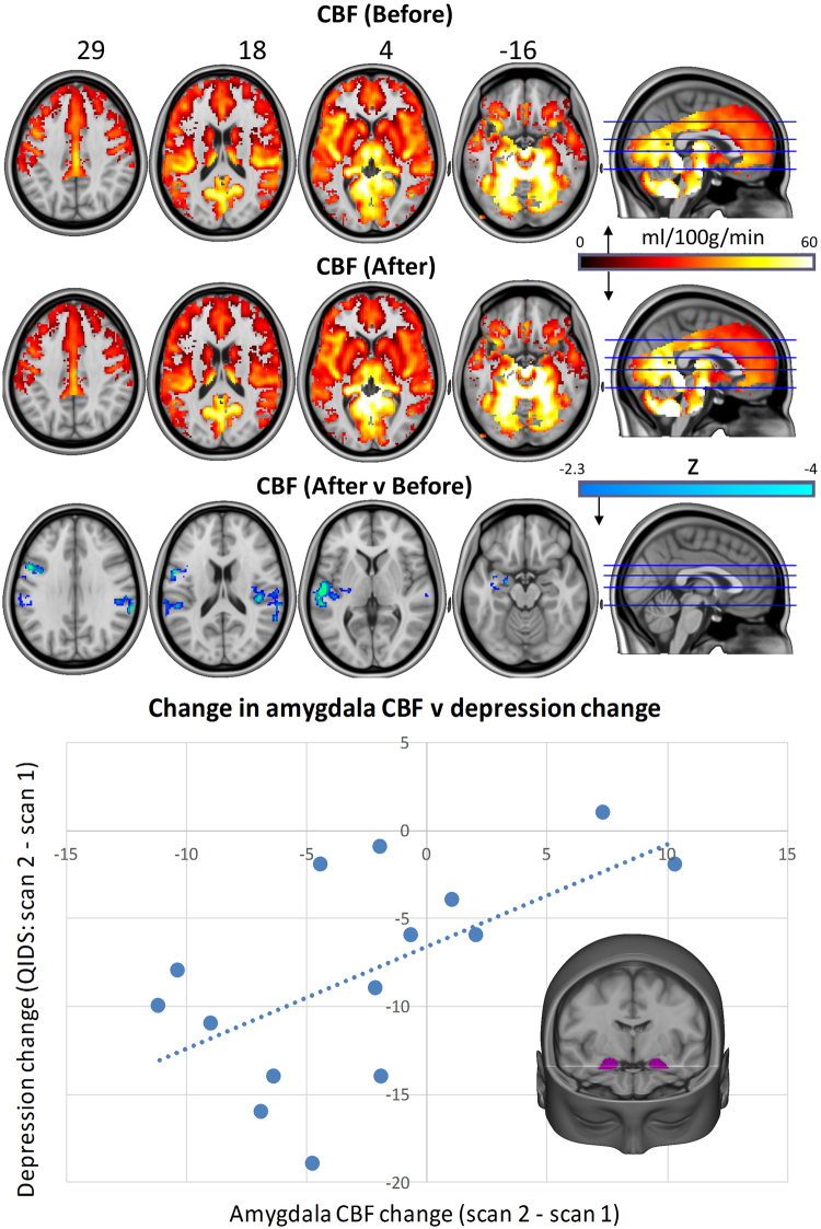
Figure 1.
Whole-brain cerebral blood flow maps for baseline versus one-day post-treatment, plus the difference map (cluster-corrected, p < 0.05, n = 16). Correlation chart shows post-Treatment changes in bilateral amygdala CBF versus changes in depressive symptoms (r = 0.59, p = 0.01). One patient failed to completed the scan 2 QIDS-SR16 rating, reducing the sample size to n = 15 for the correlation analysis. In all of the images, the left of the brain is shown on the left.

