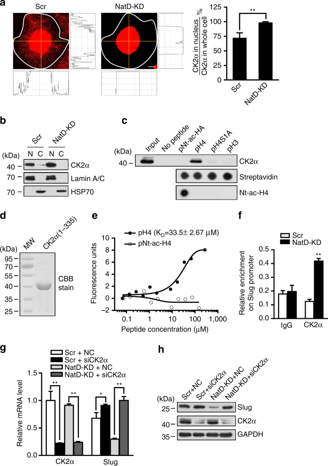
Fig. 7.
Downregulation of Nt-acetylation of histone H4 facilitates nuclear accumulation of CK2α and its binding to histone H4 in lung cancer cells. a Representative confocal scanning images of CK2α localization in Scr and H1299 cells (left panel). The staining intensity of CK2α was quantified by software ImageJ from NIH (right diagram). Scale bars, 5 μm. Results are shown as mean ± s.d. from more than 30 cells from three independent experiments; two-tailed Student’s t-test, **P < 0.01 compared with the Scr control. b Western blot analysis of CK2α distribution in scrambled and NatD-KD H1299 cells with indicated antibodies. N nucleus, C cytoplasm. c Peptide pulldown assay to detect the interaction between H4 (1–31) or H3 (1–20) peptide (pNt-ac-H4, pH4, pH4S1A, or pH3) and CK2α in H1299 cell nuclear extracts (top panel). Equal peptide biotinylated on C terminus is shown by dot blot analysis with streptavidin (middle panel). Nt-ac-H4 was confirmed by dot blot analysis with anti-Nt-ac-H4 antibody (bottom panel). d SDS-PAGE analysis of purified recombinant CK2α (1–335) from E. coli stained by Coomassie brilliant blue (CBB). MW: protein molecular weight markers. e MST assay to identify direct interactions between CK2α (1–335) and H4 or Nt-ac-H4 peptide. The dissociation constant (K D) between CK2α (1–335) and H4 peptide is 33.5 ± 2.67 μΜ. f ChIP analysis of the enrichment of CK2α on the Slug promoter in Scr and NatD-KD H1299 cells. IgG served as a negative control. Results are shown as mean ± s.d. from three independent experiments; two-tailed Student’s t-test, **P < 0.01 compared with the indicated control. g Quantitative real-time PCR analysis of mRNA levels of CK2α and Slug normalized to GAPDH in Scr and NatD-KD H1299 cells in the absence or presence of siRNA to CK2α. NC, siRNA mimics negative control. Data are mean ± s.d. of three independent experiments; Student’s t-test, *P < 0.05, **P < 0.01 compared with the indicated control. h Western blot analysis of indicated proteins from Scr and NatD-KD H1299 cells in the absence or presence of siRNA to CK2α. GAPDH served as a loading control

