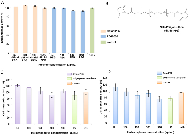
Figure 4.
Cell metabolic activity on rat cardiomyoblasts H9C2 treated with control polymers and hollow spheres. (A) Linear dithiolPEG, linear PEG2000 after 24 hours of exposure and (B) the dithiolPEG chemical structure; alamarBlue® assay was carried out on the dithiolPEG (orange), PEG2000 (blue), and untreated cells (green) (n=3, p<0.05). Cell metabolic activity on the collagen-dithiolPEG hollow spheres (C) and collagen-4armPEG hollow spheres (D) (n=3, p<0.05). Polystyrene (green) and untreated cells (orange) were used as controls

