FIG 1.
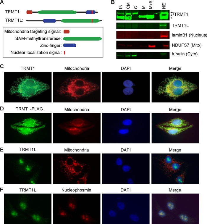
Subcellular localization of TRMT1 and TRMT1L. (A) Schematic of TRMT1 and TRMT1L with predicted domains and localization sequences. (B) TRMT1 is highly enriched in purified mitochondrial and nuclear fractions indicative of mitochondrial import and processing. IN, input; CM, cytoplasmic and mitochondrial fractions; C, cytoplasm; M, mitochondrial fraction; Mx5, 5-fold more mitochondrial equivalents loaded; NE, nuclear extract. Arrows indicate TRMT1 isoforms. *, nonspecific band detected by anti-TRMT1 antibody. Lamin B1, tubulin, and NADH:ubiquinone oxidoreductase core subunit S7 (NDUFS7) served as nuclear, cytoplasmic (Cyto), and mitochondrial (Mito) markers, respectively. (C and D) Endogenous TRMT1 and TRMT1-FLAG display nuclear and mitochondrial localization in HeLa cervical carcinoma cells. (E and F) TRMT1L is localized primarily to the nucleus and colocalizes with the nucleolar marker nucleophosmin. Mitochondria were identified using mitochondrion-targeted red fluorescent protein, and nuclear DNA was stained with DAPI.

