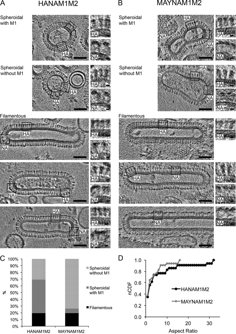
FIG 2.
Morphology of HANAM1M2 and MAYNAM1M2 particles by cryo-ET. Slices of tomograms were reconstructed from a tilt series collected using a Krios microscope at 300 kV with a Volta phase plate and nominal defocus of −0.5 μm. The slices capture spheroidal particles with and without an M1 layer and three examples of filamentous particles, isolated from supernatants of the HANAM1M2 (A) and MAYNAM1M2 (B) transfected 293T cells (scale bar, 50 nm). The M1 layer is present underneath the membrane of filamentous VLPs and in some spheroidal particles in both HANAM1M2 and MAYNAM1M2 samples. Examples of regions containing HA or NA spikes are surrounded by a black rectangle, and magnified views of the respective regions are on the right (scale bar, 5 nm). MAYNAM1M2 filamentous particles incorporate more NA spikes than HANAM1M2 filamentous particles. The MAYNAM1M2 filamentous particle in the bottom image contains only NA spikes. Two representative tomograms were deposited into the wwPDB under accession numbers EMD-8843 and EMD-8844. (C) Column plot showing percentage representation of filamentous and spheroidal particles with and without the M1 layer for HANAM1M2 and MAYNAM1M2 isolates. (D) A normalized cumulative distribution function (nCDF) plot shows that the HANAM1M2 and MAYNAM1M2 particles have similar distributions of aspect ratios (P = 0.856, Kolmogorov-Smirnov test).

