
FIG 2.
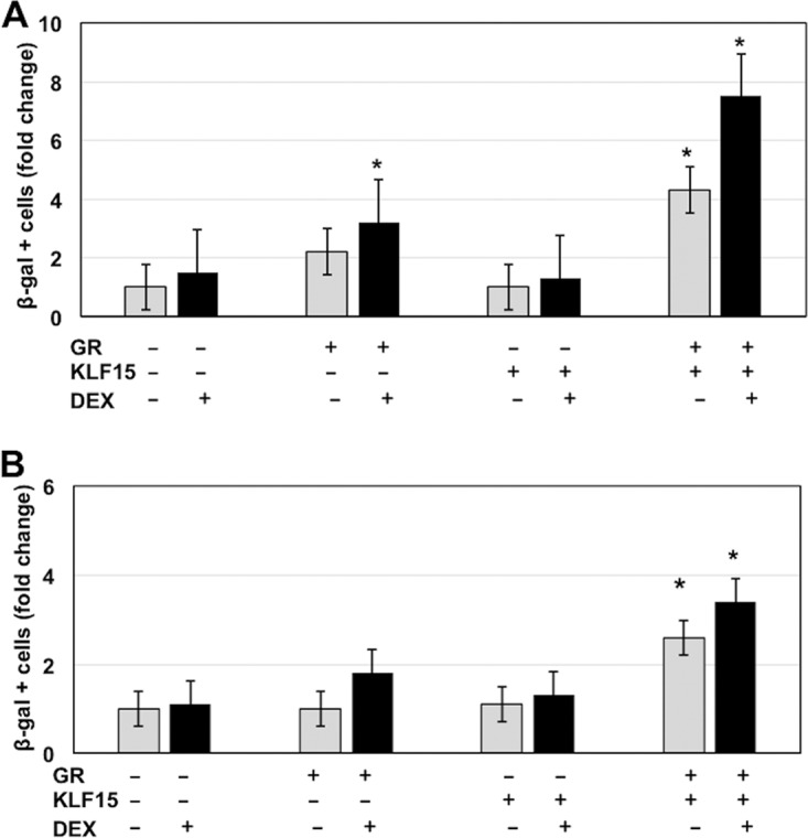
KLF15 and the GR cooperate to stimulate productive infection. Neuro-2A cells (A) or rabbit skin cells (B) were used for these studies. Twenty-four hours prior to transfection, 2% stripped fetal calf serum was added to the medium. Stripped fetal calf serum was used for these studies because normal serum contains steroid hormones, which activate the GR, as judged by nuclear localization of the GR following incubation with normal fetal calf serum (13). Cells incubated with stripped fetal calf serum for 24 h contain little or no nuclear GR. Cells were then transfected with 1.5 μg of BoHV-1 gCblue and, where indicated, a plasmid that expresses the mouse GR protein (1.0 μg DNA) and KLF15 (0.5 μg DNA). To maintain the same amount of DNA in each sample, empty vector was included in the samples. Designated cultures were then treated with water-soluble DEX (10 μM; Sigma). At 48 h after transfection, the number of β-Gal+ cells was counted. The value for the control (gCblue DNA treated with PBS after transfection) was set at 1. The results from DEX-treated cultures (black bars) were compared to those with the controls (white bars) and are an average of three independent studies. An asterisk denotes a significant difference between Neuro-2A cells transfected with BoHV-1 DNA (P < 0.05), using the Student's t test.