FIG 2.
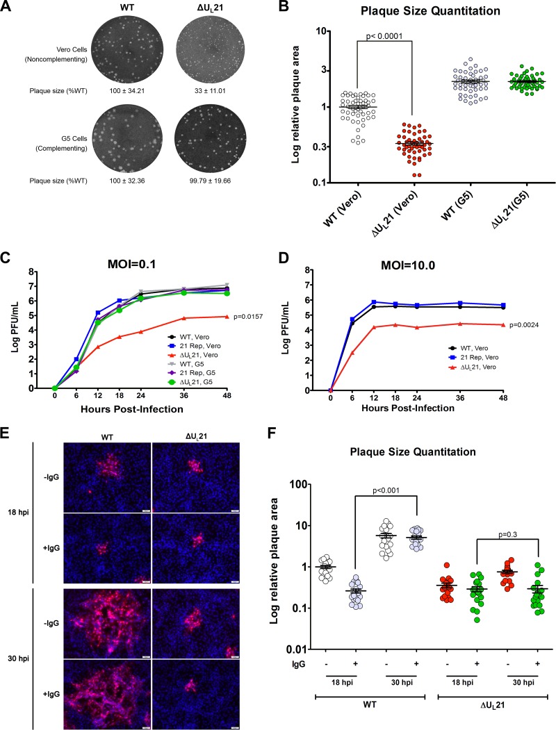
UL21 is not essential for HSV-1 replication but is needed for cell-to-cell spread. (A) Plaques of the wild-type or UL21-null mutant virus were developed on Vero or G5 cells, and representative images are shown. The areas of 50 plaques per virus type were measured using ImageJ, and the average ± standard deviation is indicated below each image, with the mean value for the wild-type sample on either cell type set to 100%. (B) Individual plaque measurements are shown. The data were graphed using a logarithmic y axis to allow for better visualization of the distribution of data points. Each circle represents an individual area measurement, and error bars show means ± standard errors of the means (SEM). (C and D) Confluent monolayers of Vero or G5 cells were infected at either a low (0.1) (C) or high (10.0) (D) MOI. At approximately 6-h intervals postinfection, total virus (cells and medium) was harvested and titrated on Vero cells. The experiment was done in duplicate, and average values are shown. (E) Vero cells seeded onto glass coverslips were infected with the WT or ΔUL21 virus at an MOI of 0.001 in the presence (+IgG) or absence (−IgG) of 5 mg/ml of human IgG to neutralize any free virus released into the medium. At 18 or 30 h postinfection (hpi), cells were fixed with paraformaldehyde, and foci of infection were immunostained with an antibody against the major capsid protein VP5 (red). Nuclei were stained with DAPI (blue). A representative image from each infection is shown. (F) The sizes of 20 plaques from the images used for the experiment described for panel E were measured. For ease of comparison, the resulting plaque area measurements were normalized, with the mean value for plaque size resulting from wild-type infection at 18 h postinfection without IgG overlay (WT, 18 hpi, −IgG) set to 1.

